You may want to supplement whatever you find in this site on the subject of Teeth Development and Function with a section on teeth in Wikipedia – they cover many important topics with good references to the literature in areas you may want to delve into more deeply.
Organization of this Chapter
You May Skip to Whatever Subject Interests You Now
Tooth Naming System
Contact with Opposing Teeth
Contact with Adjacent Teeth
The Roots
Inside the Tooth – Materials of Construction
Developmental Magic and a little Philosophy
Tooth Shapes
Man’s Best Friends
………………………………………………………………………………………………………………………
When discussing the shapes of teeth we need to look at the biting or occlusal surfaces and as well at the lateral contours which are visible toward the cheek, toward the tongue, and toward the two adjacent teeth. The contacts between adjacent teeth are important as are the contacts with opposing teeth. We will also discuss the number and shape of the roots of each tooth, because in cases of advancing periodontal (gum) disease these roots become exposed and proper home care demands a knowledge of these shapes.
Tooth Naming System
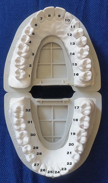
Each tooth has a letter or number assigned to it. The following diagram will be useful because it indicates the numbers for adult teeth. The common names for the TYPES of teeth are molars, premolars (or formerly bicuspids), canines (or cuspids or “eye” teeth), and incisors. The mouth is divided into 4 QUADRANTS, two on each side of top and bottom, that is there are two MAXILLARY quadrants (top), 1 through 8 and 9 through 16 and two MANDIBULAR quadrants (bottom) 17 through 24 and 25 through 32. In each quadrant there are two incisors (central and lateral, 8 and 7 for example), one canine (6 for example), two premolars ( 4 and 5 for example) and three molars (1, 2, and 3 for example). With multiple teeth of the same type they are individually termed first, second and for molars third. The third molars are more commonly called wisdom teeth.
Unusually there are other teeth that erupt in the mouth beyond the normal number, and these are called supernumerary teeth. There may be an extra premolar, for example. When I was in my 50s I had a FOURTH molar erupt behind a wisdom tooth. I was not aware of it until that time. Eventually I had it extracted more for periodontal reasons than anything else – but I kept it for 15 years, largely because I thought that if this tooth was more wise than a wisdom tooth, best to keep it around. I am happy to report that since I had it extracted, neither my wisdom nor whatever is more than that seem to have decreased more than I can tolerate.
The “corners” of the mouth are roughly marked by the canines These teeth are long and pointed, have thick roots, are very important in directing how the teeth move over each other whey you slide your teeth around, and more-or-less demarkate the division between the teeth which are prominent in your smile (anterior teeth) and those which run back deep into your mouth (posterior teeth). The reason for calling them canines, of course, relates to their pronounced size and shape in dogs (see model at end of chapter). Of course, these teeth are designed for tearing muscle fibers in the carnivore, and for biting and holding. Contrary to popular belief, these teeth are NOT designed for the opening of beer bottles, although they are admirably suited to the task!
The baby or primary teeth have letter designations in the U.S., running from A to J on the top and K through T on the bottom. Kids have the same number and types of anterior teeth as adults, but only two molars in the posterior and no premolars. The primary molars overlay the adult premolars. Note, then, that when the adult molars grow in they surface BEHIND all of the primary teeth.
Since this site is mainly concerned with adult problems, we will not address the primary dentition to a great extent until Chapter V.19, other than referring to their role in directing the eruption of permanent teeth.
Contact with Opposing Teeth
Visit Chapter II.5 for more detail on the Bite!
The normal adult dentition consists of 32 teeth, whereas the primary dentition has 20 teeth. Thus the human body produces 52 teeth, no two of which are the same. Amazingly, each unique tooth has a unique purpose! We will learn more about each tooth shape as we proceed.
The upper and lower anterior teeth can touch each other near their incisal edges (the relatively straight edge furthest from the gingiva or gum line), whereas the posterior teeth touch the opposing teeth on their OCCLLUSAL surfaces. Importantly, all teeth in the upper arch must touch some tooth in the lower arch in the normal bite, for nothing else will arrest the continuing eruption of the teeth into the mouth!

Anterior teeth with supporting aveolar bone ridge and basal bone.
The occlusal surfaces of the posterior teeth have points (cusps, like mountain peaks), ridges downward from the cusp tips, fossae (low areas between cusps), and grooves (valleys between the cusps). The more robust cusps on the upper teeth are those closer to the palate (roof of mouth), and are designated palatal cusps (or less precisely, lingual cusps – closer to the tongue). When you bite these are the cusps which fit into the fossae of the lower teeth. On the other hand, the facial cusps (toward the face or cheek) of the lower teeth are heavier and fit into the fossae of the upper teeth. These heavier cusps are normally termed the working or functional cusps due to their importance in the chewing function.
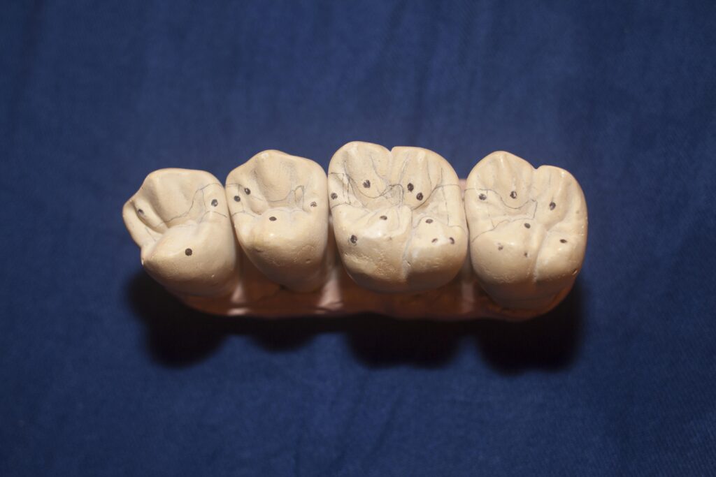
This model shows the shapes of the upper back teeth, and where they typically touch the opposing teeth.
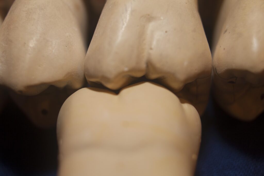
The picture of the model below shows a lower first molar such that the cusps and fossae can be visualized. In the fossae there are grooves and pits, also there are supplementary grooves running up the sides of each cusp defining ridges. These grooves are considered to have a function in defining the movement of food during chewing or in sharpening the contours where opposing teeth touch, for more efficient chewing. The grooves that extend facially and palatally define areas where opposing cusps pass during lateral movement of the jaw. It is important, when restoring a tooth that has been damaged by decay or breakage, to attempt to re-create the contours of the cusps, fossae and grooves as much as possible to fit opposing teeth, whether in stationary position or moving.

Note also, that since the functional cusps are robust, the non-functional cusps are lighter in construction. If these cusps are undermined by the restorative process, that is, their connection to the rest of the tooth substance is severed or interrupted by some filling or restorative material, then these cusps are liable to break off. A favorite offender is the popcorn “nib”, which is often encountered in movie theaters, and during action films may, due to inattention, forcefully encounter two opposing teeth in a catastrophic fashion, causing loss of a non-functional cusp. I’ve experienced this on two occasions and it is always surprising – and an expensive surprise. Best to stick to romantic comedies!
These models show how the teeth are designed to fit together.
Contact with Adjacent Teeth
It is important that each tooth contact its neighbor. If there is no contact, especially for the posterior teeth, the tooth will MOVE to make a contact.
It is worth noting at this point that your teeth never stop “wanting” to move. Forces are exerted on each tooth by the tongue, cheeks, adjacent teeth and opposing teeth, plus eruption forces. Any imbalance in these forces will cause the tooth to move, at ANY age!
The position of the contact (position from lingual to facial) and the size of the contact area are important for determining the stability of the teeth against each other and how readily cleaned is the area between the teeth. Most pairs of teeth contact each other toward the facial surfaces, meaning that the funnel shaped areas (embrasures) approaching the contact are wider on the lingual than facial. The following picture shows this geometric situation. Esthetically, the small embrasures on the facial, most visible, surfaces, particularly for the anterior teeth, seem to broaden the appearance of the teeth, and we have come to find this attractive.
There is also an embrasure toward the occlusal, it is small but important. This smooth, rounded embrasure is a consequence of a marginal ridge (adjacent teeth contact normally at the location of the marginal ridge) which is rounded and strong – often opposing teeth will contact on this marginal ridge. If the occlusal embrasure is lost when the tooth is restored incorrectly with filling material, or if the tooth is ground flat, then these edges are prone to breakage and also it is very difficult to slide dental floss past the contact to permit cleaning the sides of the teeth. This is one way in which the quality of work of the general dentist can effect adversely the periodontal condition.
Important terms which may be used here to label the two contacts that exist for each tooth are MESIAL and DISTAL. The mesial contact, on the mesial surface, is toward the midline (between the two central incisors), while the distal surface is that which is more toward the back of the mouth.
Now we have named all five surfaces of a tooth, occlusal (or incisal for the anterior teeth), facial, lingual (or palatal for the maxillary teeth), mesial and distal. Note that we can now use labels to identify any site on the entire dentition, e.g. the facial surface of tooth #14.
BUT – I usually will NOT use these technical terms in this site, for many people may be reading a section without having first looked at this chapter. So you DON’T have to memorize anything.
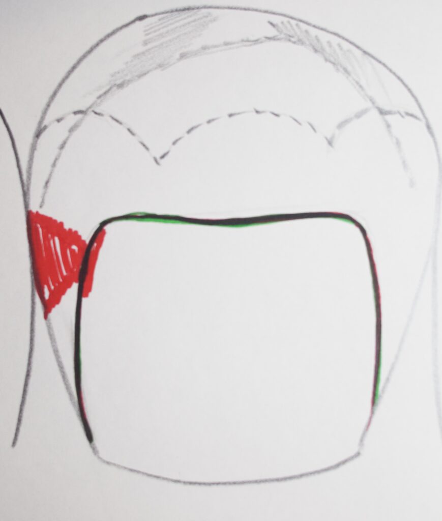
It is valuable to note that the area UNDERNEATH the proximal (with adjacent tooth) contact, between the contact and the gingiva (gum tissue) is a very common site of decay, even in the days of fluoride. The sketch below shows the area of concern (with decay in red), and it can be visualized that this part of the tooth is much less readily swept by protective saliva than other surfaces.
The Roots
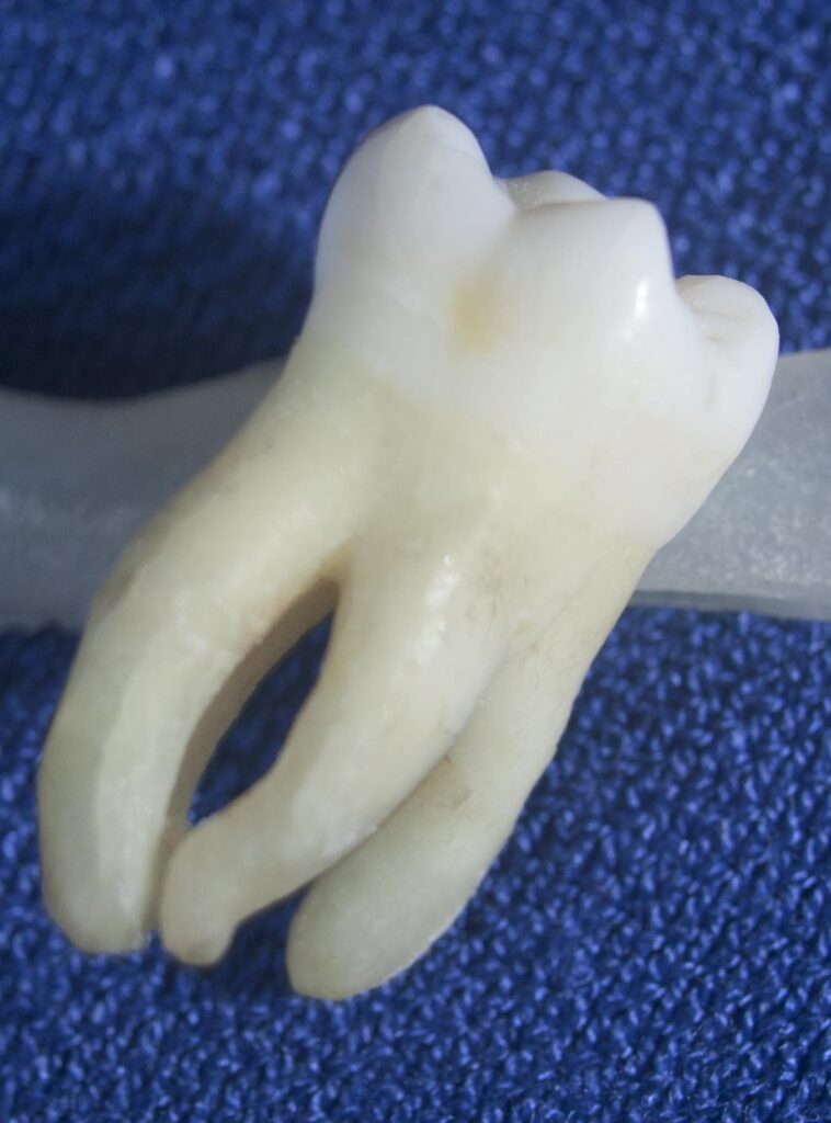
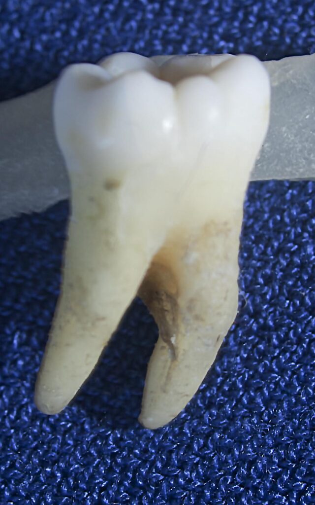
The anterior teeth will generally have only 1 root, as is true for the premolars expect for the upper first premolar, which has two roots positioned facially and palatally. The upper molars have three roots, while the lower molars have two, positioned toward the two adjacent teeth.
The pictures below show the root formations on upper and lower molars. The areas where pairs of roots come together are called furcations. These are not particularly important in the idea case, because these areas are embedded in the bone. But even moderate gum disease results in a loss in bone level and the furcations become exposed to some extent. It is not unusual that in severe cases one can pass a dental explorer (pick) or even a small brush completely through the furcations from one side of the tooth to the other! This amount of bone loss and gum recession has severe consequences, which we will describe later.
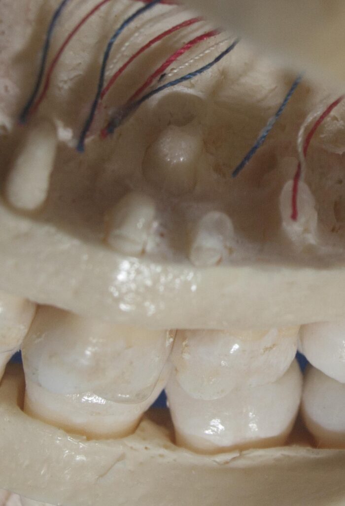
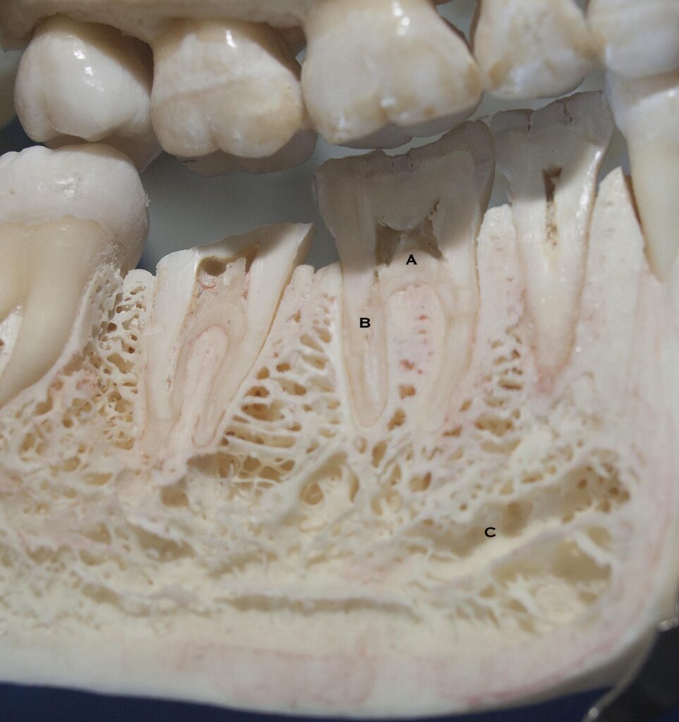
Also, in the following picture you can see a cross section of an actual human jaw, showing the roots of the teeth intact, and the pulp chamber and root canals following the root to the apex in the bone. You can also see the nerve canal in the jaw (mandible) which conducts nerves to each of the teeth.
Inside the Tooth – Materials of Construction
Chapter II.2 will go into much more detail about the internal parts of the teeth.
There are three main components of the tooth: the enamel, the dentin and the pulp. The enamel covers the part of the tooth that is exposed in the mouth in the idea case, i.e. for a teenager who has not had any bone loss or gum recession. The dentin makes up the bulk of the tooth, underlying the enamel in the crown (exposed) portion and comprising the bulk of the roots. The pulp space is where the nerves, blood vessels, lymphatic channels, immune system and cells of the tooth are located. It lies completely within the dentin and extends from the pulp chamber (within the crown of the tooth) to the root tips (the root canals).
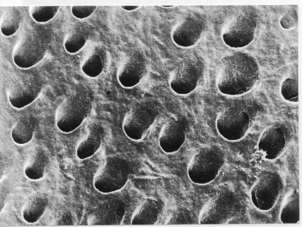
The enamel is the hardest substance in the body. It is 95% mineral and has almost negligible porosity. The dentin is, on the other hand, only 60% mineral, and is very porous. In fact, the dentin is penetrated by millions of little tunnels, called dentin tubules, which extend from the pulp chamber to the dentin-enamel junction (the DEJ). The photograph below shows these tubules in detail.
The pulp chamber contains cells called odontoblasts which line the inner dentin surface. These cells make new dentin, either slowly as part of the aging process, or more rapidly to protect the pulp from decay which is invading the tooth substance. The odontoblasts have long finger-like projections which extend through the dentin tubules to the enamel. Along with these projections there are sensory nerve fibers which run through the tubules, sensing any movement or stimulation.
The bulk of the pulp tissue exists to maintain the odontoblasts and to help ward off any infection which may invade.
Developmental Magic and a Little Philosophy
Now we know how many teeth there are and that each is unique, we know that they are all positioned in the jaw so they touch each other’s adjacent surfaces AND fit into the occlusal contours of the opposing teeth. What we do NOT know, however, is how they got there! While this subject is touched upon elsewhere in this site – it is worth mentioning here – because it is so AMAZING!
Consider that while we are developing in the womb, there are specific cells in the body from which each tooth grows. Each tooth has a particular type of cell – 52 in all. These stem cells develop first in part of the embryonic nervous system, and MIGRATE to the forming jaws.
How do these cells know where to go – so they stop in the exact position for growing the tooth it is designed to make?
We can only hypothesize that as for so much of the living organism, cells are covered with unique patterns of molecules on their surface, and when this pattern matches a complementary pattern somewhere else in the developing body, they stay there.
Cells migrate around in the blood stream, out through capillaries into surrounding tissues like bone – looking for home. In and out of the blood vessels they go, exploring, until the molecular lock-and-key is discovered. Then, they stay and grow the teeth in the programmed way. This probably happens while the bone is forming at the same time.
Now, WHAT in the bone places the specific recognition sites that the tooth-developing cells find, is a mystery to me.
In general, the more you understand about the details of the design of living systems/beings, the more you realize that there is much more that you and maybe NOBODY will ever fully understand.
This has been discovered by atomic physicists, astronomers, developmental biologists – all those whose curiosities have taken their lives to the ultimate depths we, as human beings, can achieve.
One can only speculate that there is some truth that underlies every scholarly endeavor, and that man may never know this truth – but accept its existence as a matter of faith.
Most of us in the healthcare professions are maintenance personnel and devote our lives to keeping our patient’s lives and bodies comfortable and working as well as possible. The architect? Now, that is another story entirely!
The Development of Tooth Shapes
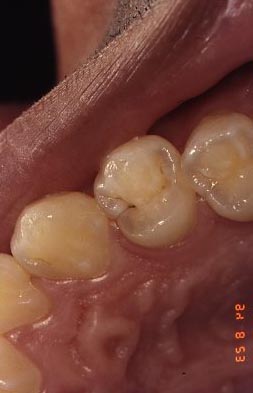
For the last consideration in this section, it is worth thinking about how the particular shapes of each tooth develop. I’ve referred to cells that migrate to places in the bone, and that these cells divide into each type of tissue required for tooth development. These are, in essence, stem cells – which can divide into different types of cells – some of which form dentin, enamel, the cementum covering of the root, the pulp tissues, etc. There are also regions of the tooth that develop largely independently – these are called DEVELOPMENTAL LOBES. For example, for a upper first premolar, there are four separate and distinct developmental lobes – three for the facial cusp and one for the lingual/palatal. Each developmental lobe forms a part of the tooth that is largely spherically symmetrical. But, since the facial cusp is formed from three lobes, and these can be different sizes and heights, the cusp can have a rather complex shape, involving indentations and ridges. The lingual cusp, however, develops from only one lobe, so it is largely rounded and symmetrical.
The picture of the patient’s upper first premolar above illustrates that point perfectly.
Man’s best Friends
These are models that are used by student animal doctors to study their teeth and practice maintenance.
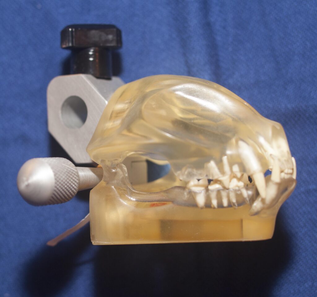
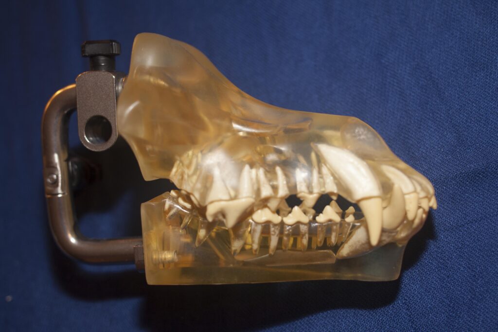
Models of cat and dog dentition. Students invariably think they are of dangerous animals!